MPC1 is expressed in the human hair follicle and MPC inhibition blocks cell cycle progression and disrupts the expression of hair follicle signaling pathway genes. A) MPC1 and PDK immunoreactivity in human hair follicles. CTS connective tissue sheath. DP – dermal papilla; DSC – dermal sheath cup; GL–germ layer; HS – hair shaft; IRS – inner root sheath; L-ORS – lower outer root sheath; SG – sebaceous gland. Regional analysis (analysis zones indicated by red dotted lines) performed on 7 anagen hair follicles from 3 donors. Mann Whitney test, p-value *** 0.0006. Scale bar 50 µm. B) Fluorescent EdU labeling on human hair follicle tissue sections shows how treatment with UK-5099 blocks DNA replication in the hair follicle, both in the bulging epithelium and in the hair matrix (HM). DP – dermal papilla. Scale bar 50 µm. C) Quantitative analysis of EdU and Ki-67 in the bulge and hair matrix after treatment with UK-5099. Ordinary one-way ANOVA with multiple comparisons. EdU analyses: adjusted p-values *** 0.0002, **** < 0.0001. Ki-67 analyses: adjusted p-values ** 0.0018; *** 0.0006; **** <0.0001. N = 2–3 donors (6–10 independent anagen hair follicles per condition). The drawn line is the average. D) Dot plot of the top 10 enriched IPA pathways after treatment with 40 µM UK-5099. Analysis performed on 1206 genes with a 2-fold change and padj < 0.05. See also S3 Fig in S1 File. E) Volcano plot annotated with differentially expressed genes involved in FGF, IGF, TGFβ and Wnt signaling with an adjusted p-value <0.05 after treatment of human hair follicles with 40 µM UK-5099. Credit: PLOS ONE (2024). DOI: 10.1371/journal.pone.0303742
× close to
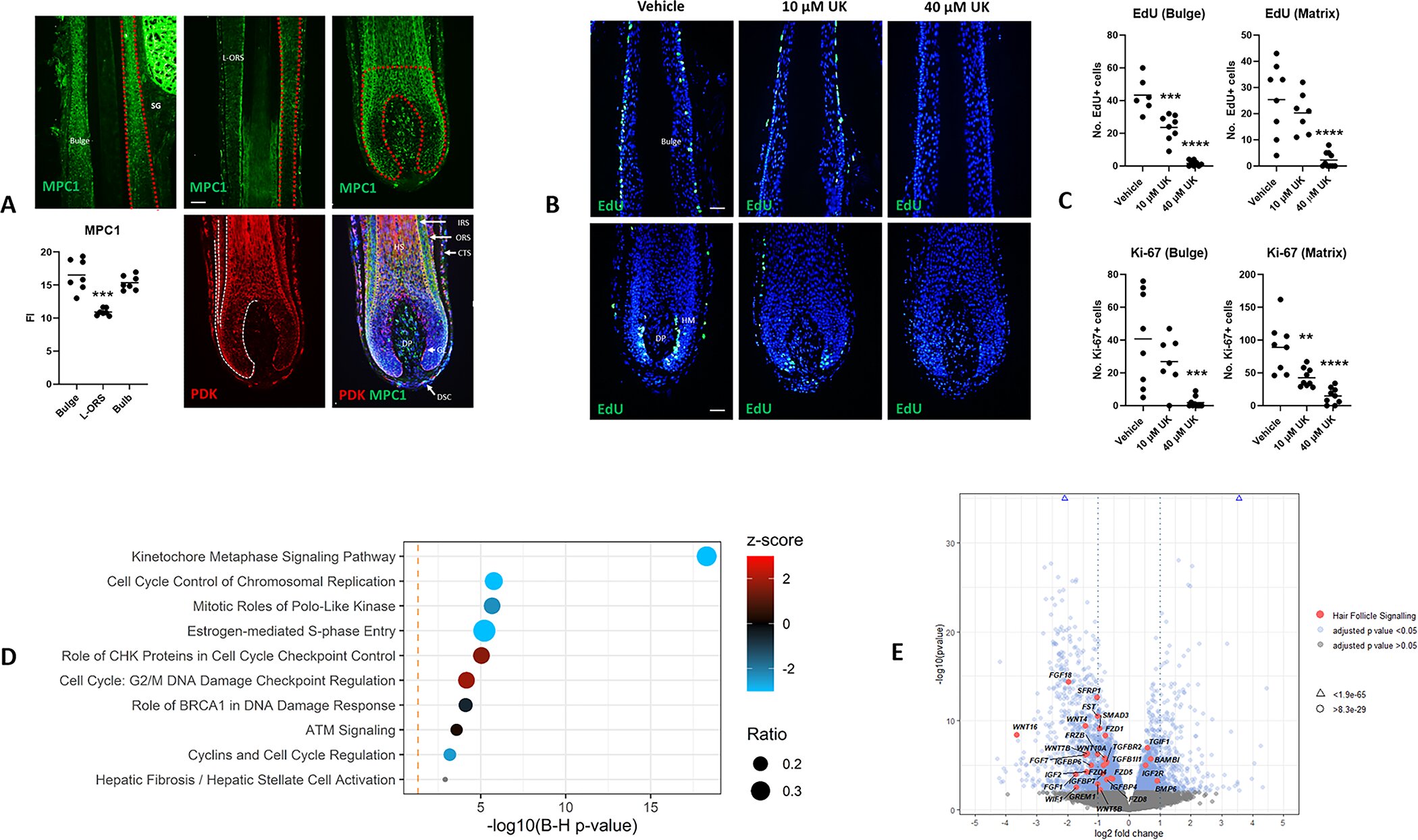
MPC1 is expressed in the human hair follicle and MPC inhibition blocks cell cycle progression and disrupts the expression of hair follicle signaling pathway genes. A) MPC1 and PDK immunoreactivity in human hair follicles. CTS connective tissue sheath. DP – dermal papilla; DSC – dermal sheath cup; GL–germ layer; HS – hair shaft; IRS – inner root sheath; L-ORS – lower outer root sheath; SG – sebaceous gland. Regional analysis (analysis zones indicated by red dotted lines) performed on 7 anagen hair follicles from 3 donors. Mann Whitney test, p-value *** 0.0006. Scale bar 50 µm. B) Fluorescent EdU labeling on human hair follicle tissue sections shows how treatment with UK-5099 blocks DNA replication in the hair follicle, both in the bulging epithelium and in the hair matrix (HM). DP – dermal papilla. Scale bar 50 µm. C) Quantitative analysis of EdU and Ki-67 in the bulge and hair matrix after treatment with UK-5099. Ordinary one-way ANOVA with multiple comparisons. EdU analyses: adjusted p-values *** 0.0002, **** < 0.0001. Ki-67 analyses: adjusted p-values ** 0.0018; *** 0.0006; **** <0.0001. N = 2–3 donors (6–10 independent anagen hair follicles per condition). The drawn line is the average. D) Dot plot of the top 10 enriched IPA pathways after treatment with 40 µM UK-5099. Analysis performed on 1206 genes with a 2-fold change and padj < 0.05. See also S3 Fig in S1 File. E) Volcano plot annotated with differentially expressed genes involved in FGF, IGF, TGFβ and Wnt signaling with an adjusted p-value <0.05 after treatment of human hair follicles with 40 µM UK-5099. Credit: PLOS ONE (2024). DOI: 10.1371/journal.pone.0303742
Scientists from the University of Manchester have linked one of the ways in which cells respond to stressful conditions to limited healthy hair growth.
The team at Manchester Hair Research Group unexpectedly discovered the link in a laboratory experiment where they tested a drug to see if it would grow hair follicles on the human scalp in a dish. The study inadvertently led to a connection with the cellular stress response – an ancient biological mechanism that occurs throughout life, from yeast and roundworms to humans.
The research was published in PLOS ONE.
The team hopes that their work in targeting this pathway could one day lead to treatments for hair loss.
Fully known as the Integrated Stress Response (ISR), it is activated during stressful cellular conditions such as poor nutrient availability, viral infections or when there is a build-up of misshapen proteins in cells. The ISR allows cells to slow down regular activities by producing fewer new proteins, causing them to enter a partial standstill to adapt and cope with the stress. However, if it doesn’t work, it can cause cells to die.
ISR is already a topic of great interest to scientists studying cancer, neurodegenerative diseases and aging.
Dr. Talveen Purba, Research Fellow at the University of Manchester and senior author of the study, said: “We were testing a drug that targets metabolism in human hair follicles to influence how cells generate energy, which – based on the work from others – we expected to stimulate stem cells, but we found the opposite was true: hair growth was instead blocked as cells, including stem cells, quickly stopped dividing.”
They also found signs that mitochondria were not functioning properly and that there were disruptions in the way cells communicate with each other. Using a combination of experimental approaches to look more closely, the team found signs that ISR activation was the cause.
Derek Pye, lead technician of the research group and co-author of the study, said: “When we look at hair follicles under the microscope, it is striking how consistent the response is between hair follicles from different people.”
Following on from this early-stage research, the team now wants to better understand the broader implications of the ISR on the hair follicle and investigate its activity in people with hair loss.
Dr. Purba added: “We are incredibly hopeful because we believe that activation of this pathway could play an important biological role in limiting hair growth in people with hair loss, meaning that targeting it could lead to new treatments. “
More information:
Derek Pye et al, Activation of the integrated stress response in human hair follicles, PLOS ONE (2024). DOI: 10.1371/journal.pone.0303742
Magazine information:
PLoS ONE