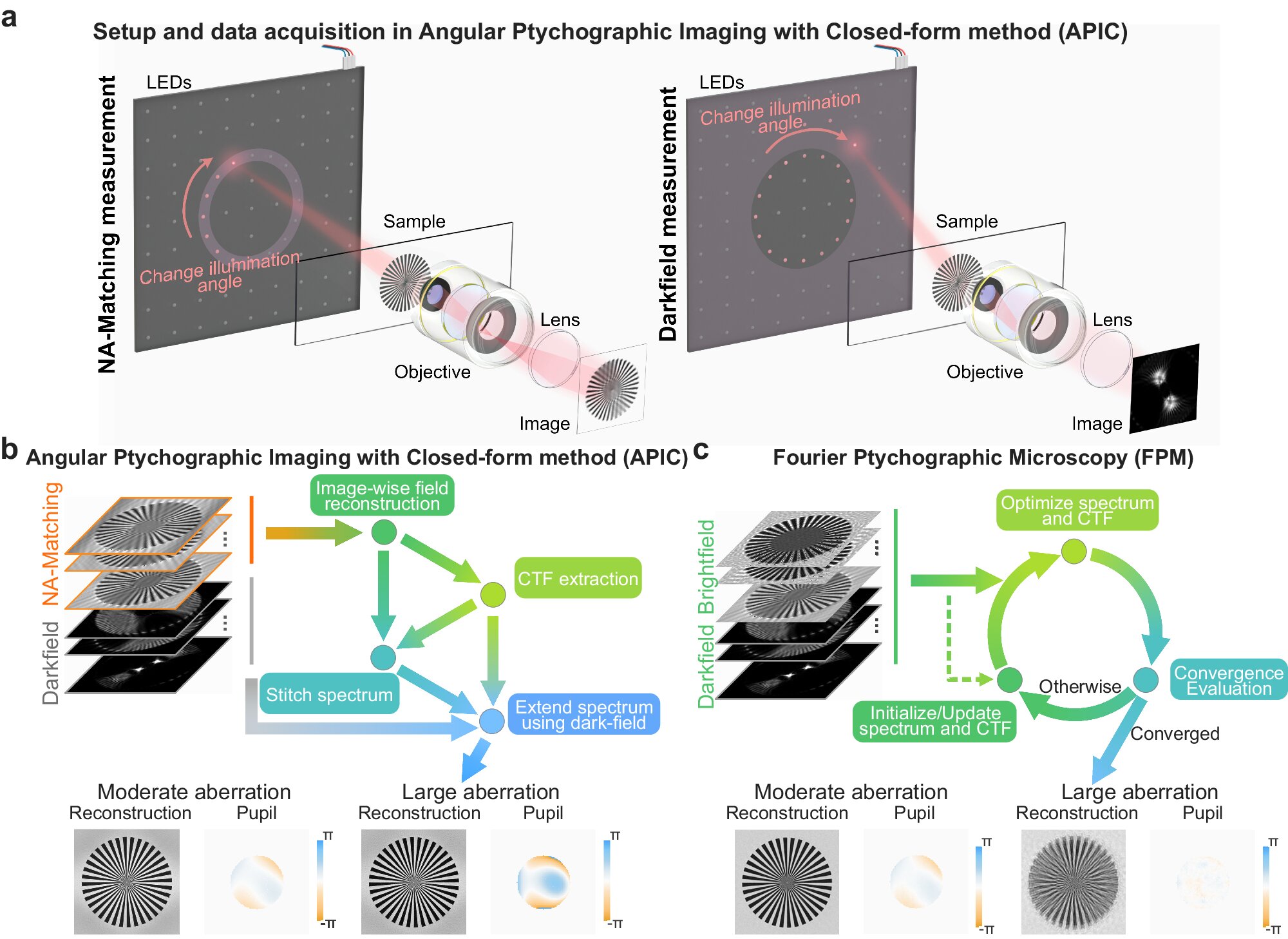
Concept of angular ptychographic imaging with closed-form method (APIC) and comparison between the reconstruction process of APIC and Fourier ptychographic microscopy (FPM). Credit: Nature communication (2024). DOI: 10.1038/s41467-024-49126-y
For hundreds of years, the brightness and magnification of microscopes were ultimately limited by the physical properties of their optical lenses. Microscope makers pushed those limits by making increasingly complex and expensive stacks of lens elements. Yet scientists had to choose between high resolution and a small field of view on one hand, or low resolution and a large field of view on the other.
In 2013, a team of Caltech engineers introduced a microscopy technique called FPM (for Fourier ptychographic microscopy). This technology marked the advent of computational microscopy, the use of techniques that combine the detection of conventional microscopes with computer algorithms that process detected information in new ways to create deeper, sharper images that cover larger areas. FPM has since been widely adopted for its ability to obtain high-resolution images of samples while maintaining a large field of view using relatively inexpensive equipment.
Now the same laboratory has developed a new method that outperforms FPM in its ability to acquire images without blur or distortion, even while taking fewer measurements. The new technique, described in an article that appeared in the magazine Nature communicationcould lead to advances in areas such as biomedical imaging, digital pathology and drug screening.
The new method, called APIC (for Angular Ptychographic Imaging with Closed-form method), has all the advantages of FPM without what could be described as its biggest weakness: namely that the FPM algorithm, to arrive at a final image, relies on starting with one or more best guesses and then adjusting bit by bit to arrive at the “optimal” solution, which may not always be true to the original view.
Led by Changhuei Yang, the Thomas G. Myers Professor of Electrical, Bioengineering, and Medical Engineering and a researcher at the Heritage Medical Research Institute, the Caltech team realized that it was possible to eliminate this iterative nature of the algorithm.
Rather than relying on trial and error to arrive at a solution, APIC solves a linear equation, which yields details of the aberrations or distortions introduced by a microscope’s optical system. Once the aberrations are known, the system can correct them, essentially performing as if this were ideal, delivering clear images covering a large field of view.
“We solve the complex field at high resolution in a closed way, because we now have a deeper understanding of what a microscope captures, what we already know and what we really need to figure out, so we don’t need iteration,” says Ruizhi Cao, co-lead author of the paper, a former doctoral student in Yang’s lab and now a postdoctoral researcher at UC Berkeley. “This way we can basically guarantee that we see the true final details of a sample.”
As with FPM, the new method measures not only the intensity of light seen through the microscope, but also an important property of light, called ‘phase’, which is related to the distance light travels. This property is not noticeable to the human eye, but contains information that is very useful in correcting aberrations.
In solving this phase information, FPM relied on a trial-and-error method, explains Cheng Shen, one of the lead authors of the APIC paper, who also performed the work in Yang’s lab and is now a computer algorithm engineer vision is at Apple.
“We have proven that our method gives you an analytical solution and in a much simpler way. It is faster, more accurate and uses deep insights into the optical system,” says Shen.
In addition to eliminating the iterative nature of the phase-resolving algorithm, the new technique also allows researchers to collect clear images over a large field of view without repeatedly refocusing the microscope. With FPM, if the height of the sample were to vary even a few tens of microns from one section to another, the person using the microscope would have to refocus for the algorithm to work.
Because these computational microscopy techniques often involve stitching together more than 100 lower resolution images to compose the larger field of view, APIC can make the process much faster and avoid the potential introduction of human error in many steps.
“We have developed a framework to correct the aberrations and also improve the resolution,” Cao says. “These two possibilities can potentially be fruitful for a wider range of imaging systems.”
Yang says the development of APIC is critical to the broader scope of work his lab is currently conducting to optimize image data input for artificial intelligence (AI) applications.
“Recently, my laboratory demonstrated that AI can outperform expert pathologists in predicting metastatic progression from simple histopathology slides of lung cancer patients,” says Yang. “That predictive ability is highly dependent on achieving uniform focus and high-quality microscopy images, something for which APIC is well suited.”
More information:
Ruizhi Cao et al, High-resolution, large field-of-view label-free imaging via aberration-corrected, closed-form complex field reconstruction, Nature communication (2024). DOI: 10.1038/s41467-024-49126-y
Offered by the California Institute of Technology
Quote: New computational microscopy technique offers a more direct route to crisp images (2024, June 28), retrieved June 29, 2024, from https://phys.org/news/2024-06-microscopy-technique-route-crisp-images.html
This document is protected by copyright. Except for fair dealing for the purpose of private study or research, no part may be reproduced without written permission. The contents are provided for information purposes only.